
Overview of the toxicological and biological analytical infrastructure
SEON has a variety of state-of-the-art and high-quality measuring instruments at its disposal to characterize a wide range of particles in detail with regard to their chemical, physical and biological properties. We offer our cooperation partners and external customers to perform corresponding analyses.
You are welcome to contact us in this regard
On this page you will find a detailed listing of our biological analytical infrastructure as well as an overview of our common analytical methods:

Photo: David Hartfiel
Beckman Coulter Gallios
Flow cytometer
In a flow cytometer, blank or stained sample cells of a cell suspension are passed through a narrow channel and separated. In the process, they are excited with lasers of different colors and a series of sensors detect the various types of light refracted or emitted by the cells. The data collected by the sensors is collected and integrated using Kaluza software to provide a comprehensive picture of the sample.
Our instrument is equipped with a sample changer and 3 excitation lasers (violet: 405 nm; blue: 488 nm; red: 638 nm). The emission of the cells is detected by 10 photomultupliers depending on the excitation laser (violet: 430 nm/40 nm BP, 550 nm/40 nm BP; blue: 525 nm/40 nm BP, 575 nm/30 nm BP, 620 nm/20 nm BP, 695 nm/30 nm BP, 755 nm LP; red: 675 nm/20 nm BP, 725 nm/20 nm BP, 755 nm LP).
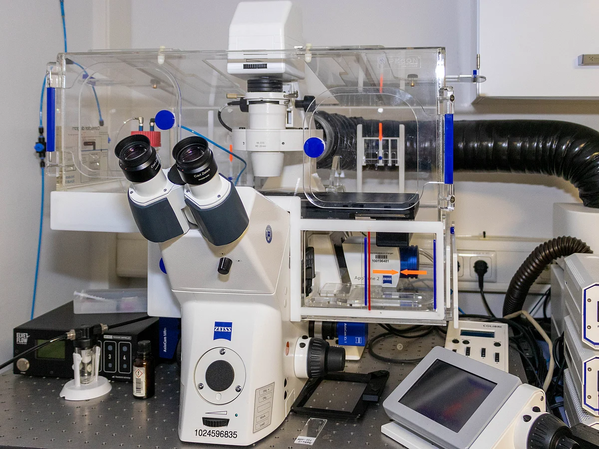
Photo: Harald Unterweger\Uni-Klinikum Erlangen
Zeiss Axio Observer
Live cell imaging Unit
Our Axio Observer fluorescence microscope from Zeiss is equipped with an incubation chamber with temperature and CO2 control. This enables real-time imaging of cells in a native incubation environment. The high-resolution digital microscopy camera also allows imaging of even fine cell structures.
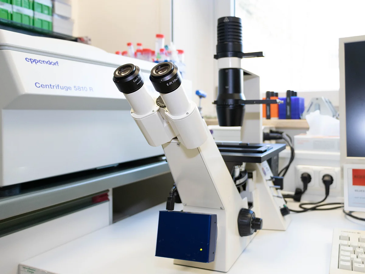
Photo: Harald Unterweger\Uni-Klinikum Erlangen
Zeiss Axiovert 40
Inverted microscope
The Axiovert 40 from Zeiss is a universally applicable inverted microscope for various applications in biology and medicine for examining cell cultures, blood or tissue samples. Various microscopy or contrast methods are available: Brightfield, darkfield, phase contrast and polarization contrast.

Photo: Harald Unterweger\Uni-Klinikum Erlangen
ProXima
Documentation system for gels
The ProXima from Isogen Life Science is a high-performance documentation system for the applications chemiluminescence, bioluminescence, UV/VIS fluorescence or densitometry of colorimetrically stained gels and blots.

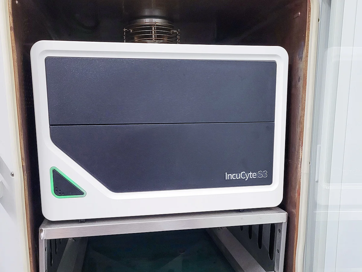
Photo: Harald Unterweger\Uni-Klinikum Erlangen
IncuCyte S3
Automated real-time cell imaging
With the IncuCyte it is possible to obtain deeper and physiologically relevant information about cells in real time. Here, kinetic data is generated by the automated acquisition and analysis of images in periods ranging from a few hours to weeks.

Photo: Harald Unterweger\Uni-Klinikum Erlangen
xCELLigence RTCA SP
Real-time cell vitality analysis
The xCELLigence system from ACEA Biosciences is a microelectronic biosensor system for cell assays. It enables dynamic, label-free cell analysis in real time for various research applications. For this purpose, specially designed microplates with interdigitated gold microelectrodes are used for non-invasive monitoring of cell viability using electrical impedance as a measurement parameter. Continuous monitoring of cell viability enables differentiation of various cell viability disorders such as senescence, cell toxicity and proliferation impairment.
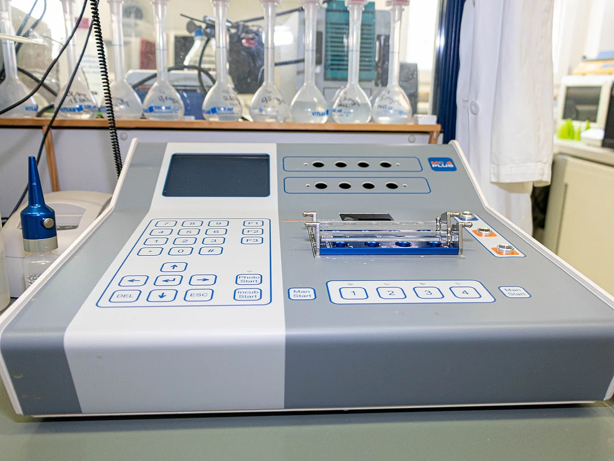
Photo: Harald Unterweger\Uni-Klinikum Erlangen
MC 4 Plus
Coagulometer
The MC 4 plus is a semi-automatic 4-channel ball coagulometer for the laboratory with medium sample volumes with mechanical detection. This device is used, among other things, to determine the coagulation time, whereby the measurement is started either manually by pressing a button or comfortably and reproducibly by an automatic start pipette connected to the device.

Photo: Uni-Klinikum Erlangen
3D Cell Explorer
3D Microscope
Nanolive's 3D Cell Explorer captures the cell's 3D refractive index distribution without labeling. The extremely low light power used to generate the holograms allows for the complete absence of phototoxicity, resulting in excellent temporal resolution.
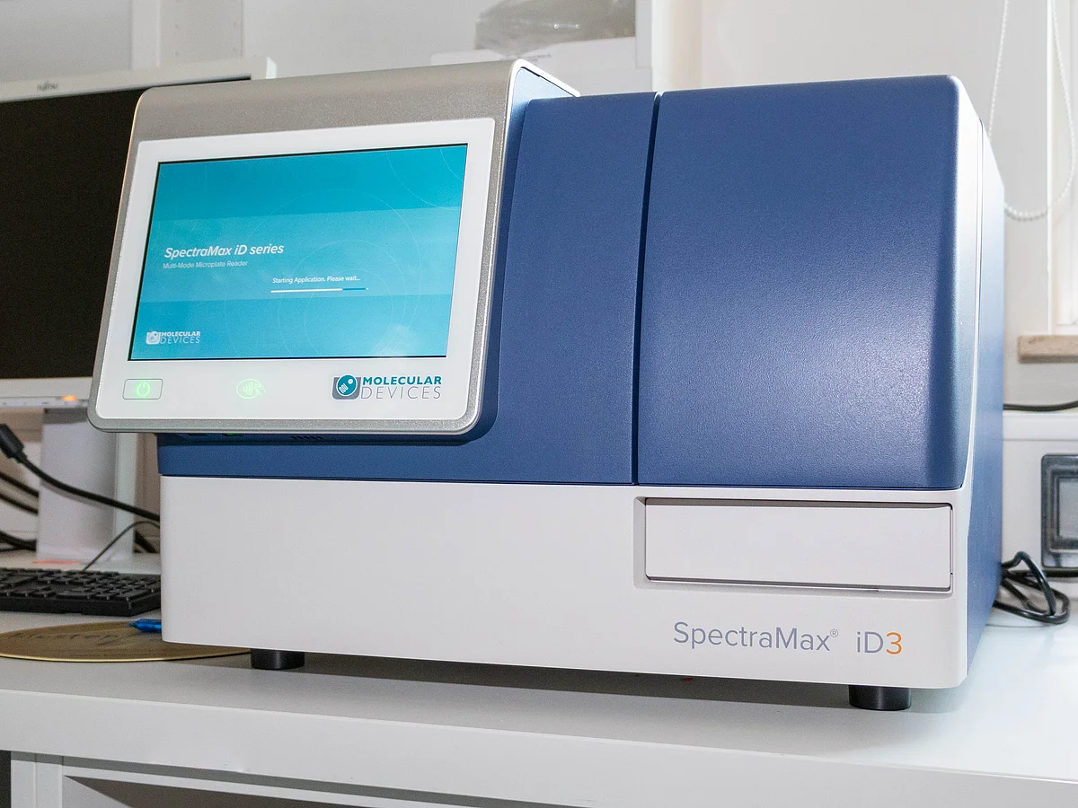
Foto: Harald Unterweger\Uniklinikum Erlangen
UV/VIS
Photometry
For direct quantification of chromophores, e.g. for various cell viability assays based on fluorescent dyes, we use a SpectraMax iD3 plate photometer from Molecular Devices.
Histology
Equipment
- Paraffin pouring station EG 1150H from Leica: heated, with microprocessor control. The large heated work surface with integrated cooling spot has a kerosene drain and one heated bath each for embedding molds and cassettes.
- Leica RM 2255 rotary microtome
- Fully automatic stand cryostat MNT from Slee. It features simple and intuitive operation, a fully automated rotary microtome, and a space-saving design. The large stainless steel chamber can be temperature controlled down to -35 °C in a very short time.
Histology
Assays (selection)
Cryo-fixation /paraffin embedding and preparation of thin sections
Staining methods
- Prussian blue
- Hematoxilin-Eosin
- Masson Goldner Trichrome Stain
- DAPI
- Characterization of cellular components by immunofluorescence staining (LAMP-1, F-actin, GM 130, EGFR, transferin receptor CD 71, ...)
Cyto-/Genotoxicity
Assay overview (selection)
- Proliferation, morphological cell analysis (real-time cell imaging: IncuCyte)
- Plasma membrane integrity (LDH, photometer)
- Cell proliferation (BrdU, photometer, flow cytometer)
- Caspase-3/7 activation (flow cytometer)
- Metabolic cellular activity (MTT, WST, XTT, photometer)
- Apoptosis/necrosis (annexin V/propidium iodide staining, flow cytometer)
- Mitochondrial membrane potential (DiIC1(5), flow cytometer)
- Cell cycle and DNA degradation (propidium iodide triton, flow cytometer)
- Calcium influx (Fluo-4, flow cytometer)
- ATP quantification (CellTiter-Glo, photometer)
- DNA double strand break (yH2AX, Western blot, flow cytometer)
- DNA damage (COMET assay, fluorescence microscopy)
- HET-CAM Assay
Oxidative stress
Assays (selection)
- intracellular H2O2 (DCFH, flow cytometer)
- Intracellular O2- (hydroethidium, flow cytometer)
- Measurement of reduced glutathione (monobromobimanes, flow cytometer)
- Membrane peroxidation (BODIPY 581/591 C11, flow cytometer)
- Lipid peroxidation (determination of malondialdehyde with thiobarbituric acid, photometer)


